-768x677.jpg "Development of Video Microscopes 1")
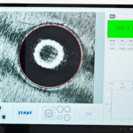
Video microscopes break through the limitations of traditional optical microscopes by integrating optical imaging and digital technology, with core advantages reflected in three breakthroughs:
- Eyepiece-Free Visualization: High-definition cameras (mostly CMOS/CCD sensors with 1920×1080 resolution) capture optical images and transmit them to a display in real time, enabling simultaneous observation by multiple users and eliminating eye fatigue from single-eye viewing. Combining optical and digital magnification, mainstream models offer continuous adjustment from 3× to 500×, with high-end products reaching up to 2000×, balancing macroscopic field of view and microscopic details.
- Intelligent Image Processing: Equipped with dedicated software for image comparison, 2D/3D measurement, defect marking, etc. Some models feature built-in AI recognition systems that automatically count defects and classify samples, improving inspection efficiency by more than 12 times compared with manual work. Paired with Z-stacking technology, the depth of field is 3–5 times that of traditional microscopes, clearly displaying concave-convex structures without complex sample preparation.
- Multi-Functional Integrated Design: Integrates HDMI/USB interfaces, ring LED light sources, laser positioners, etc. Some models support tilted observation (multi-angle sample inspection) and wireless transmission, with ergonomic design to reduce long-term operation strain. Industrial-grade products also feature ESD protection for precision electronic inspection.
-300x264.jpg "Development of Video Microscopes 2")
Video microscopes have expanded from laboratories to various industries, becoming core tools for inspection and research:
- Industrial Quality Control: Inspect PCB solder joints and semiconductor wafer scratches in electronics; detect micro-cracks on engine parts in automotive manufacturing; observe precision structures in watch repair and mold processing. Some devices support DXF drawing overlay for precise positioning.
- Medical & Scientific Research: Observe blood smears and skin tissues in primary hospitals to assist anemia and fungal infection diagnosis; record dynamic processes such as cell division and crystal growth in life sciences, supporting remote consultation and data sharing.
- Education & Popularization: Display pollen patterns and paramecium movement in classrooms; popular science creators capture footage of salt crystallization and insect compound eyes, with short videos gaining over 10 million views.
- Special Fields: Analyze trace evidence in forensic investigations; observe material details during cultural relic restoration; identify microscopic features in jewelry appraisal and document anti-counterfeiting.
-2-300x137.jpg "Development of Video Microscopes 3")
Classification, Selection & Future Trends
Classified by structure and function: trinocular/monocular, large field/standard field, manual/autofocus, as well as 3D ultra-depth of field, universal bracket and other specialized models for different scenarios.
Selection Criteria
- Performance Matching: For precision samples, prioritize models with optical magnification and resolution ≤1μm; for mobile inspection, consider lightweight (≤500g) and battery life.
- Scenario Adaptation: Dustproof and shockproof for industrial sites; antibacterial design for medical use; easy operation and data sharing for education.
- Function Screening: Autofocus and parameter memory for batch inspection; image stitching and metrological traceability (compliant with ISO/IEC17025) for scientific research.
-150x150.jpg)